右室流出路起源心室期外収縮(RVOT PVC)
右室流出路起源心室期外収縮とは、右室流出路から発生する心室期外収縮のことです。日常臨床で最も頻繁に遭遇するタイプで、基礎心疾患がない特発性の場合が多く、良性とされることがほとんどです。右室流出路は心筋と肺動脈の移行部にあたり、この領域は構造的に心収縮の影響を受けやすく、発生学的に他の心室筋とは異なる性質を持つため、異常興奮が起きやすいと考えられています。
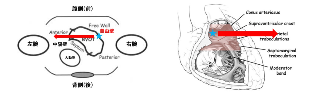
起源の分類
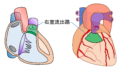
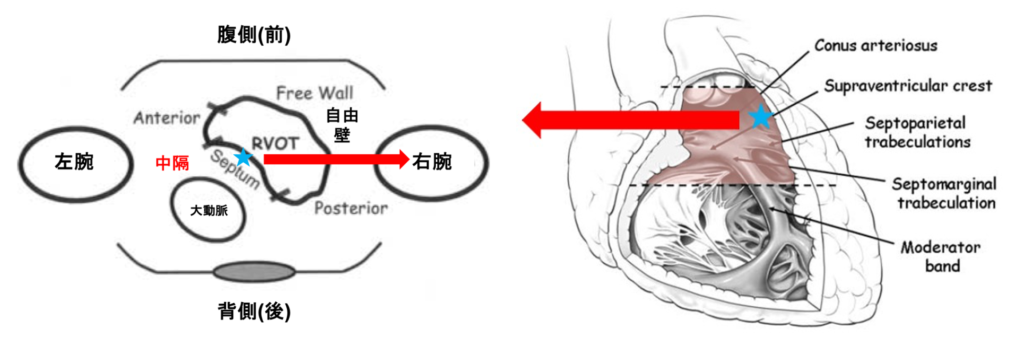
右室流出路は自由壁・前壁・後壁・中隔の4つのセグメントに分類されます。水平面における断面では三日月型をしており、腹側に自由壁、背側に中隔が位置しています。前壁・後壁は前後というよりは左右に近く、前壁は左側、後壁は右側に位置しています。12誘導心電図の所見から右室流出路の中でも4つのセグメントのうちどこを起源としているかをさらに推定することができます。

心電図所見
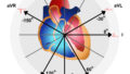
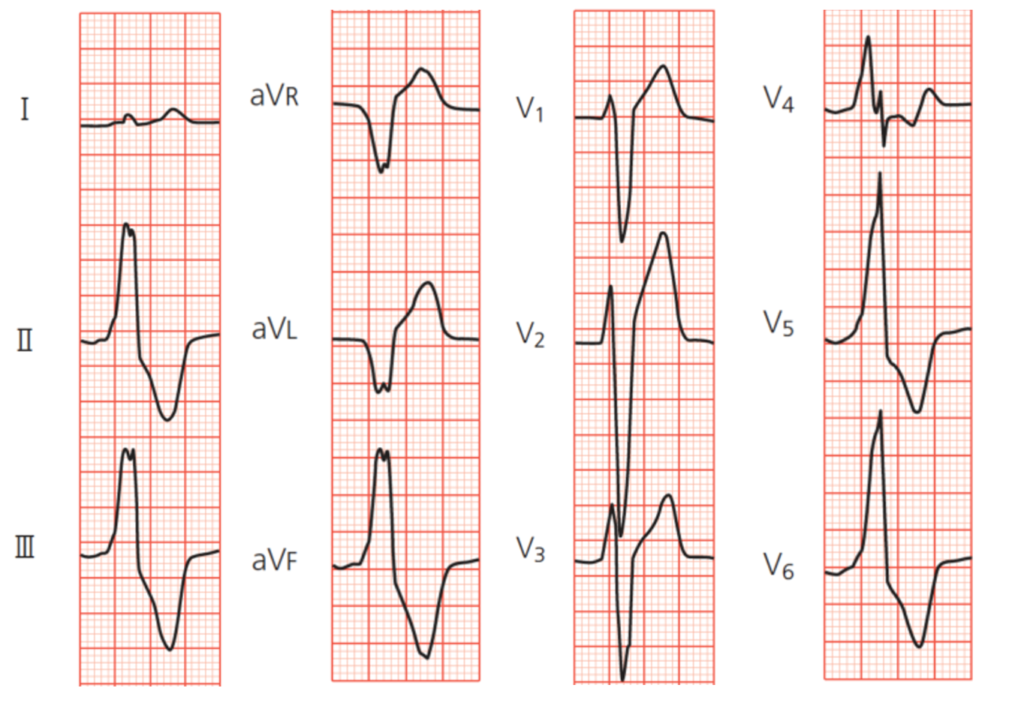
右室流出路は右室の中で最も高い位置にあり、期外収縮の興奮がそこからほとんど真下方向に向かって伝導するため下方軸となり、下壁誘導で高いR波になります。上方から見ているaVR誘導、aVL誘導では興奮が離れていく方向になるのでQS波になります。
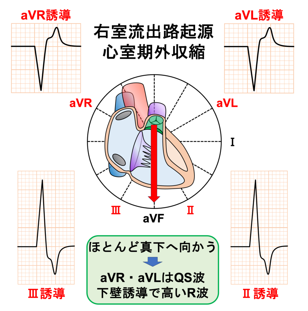
また、右室が最初に興奮してから左室に興奮が伝導するので左脚ブロックパターンになります。
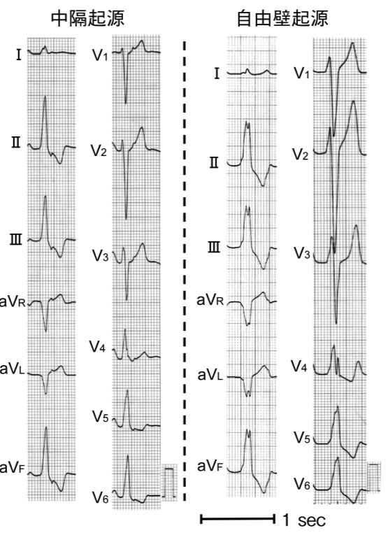
右室流出路中隔
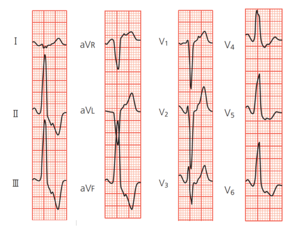
・胸部誘導の移行帯はV3~V4以降
・Ⅰ誘導で陽性成分が小さく、時に深いS波を認める
・aVL誘導とaVR誘導のQ波はaVL誘導のほうが深い
下壁誘導
中隔起源の期外収縮では、右室と左室はほぼ同時に興奮します。そのため下壁誘導でnotchのない高いR波となります
Ⅰ誘導
中隔壁は右室流出路の中で左側に位置しています。そのため、左から右に興奮が伝わります。四肢誘導で考えると、Ⅰ誘導と反対方向に向かいます。そのため、Ⅰ誘導では陽性成分が小さく、時に深いS波を認めます
aVR・aVL誘導
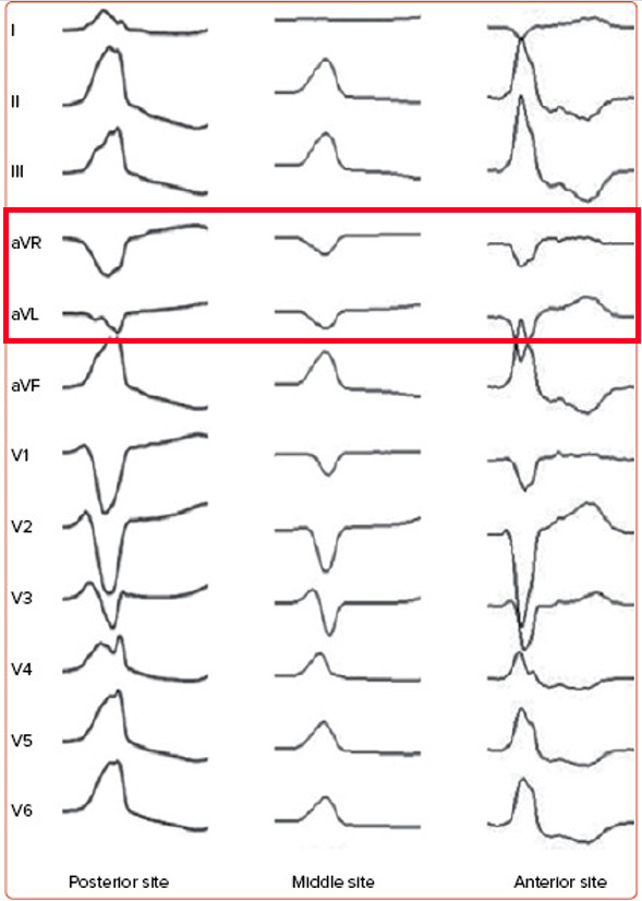
aVR誘導のQ波の振幅は、右心室流出路の前壁から後壁に向かって徐々に大きくなり、逆にaVL誘導のQ波の振幅は、右心室流出路の前壁より後壁で小さくなります。(図1)
・aVL誘導のQ波の振幅:前壁 大きい → 後壁 小さい
これは、右心室流出路が左心室流出路を包み込むようにして大動脈起始部の前側方に位置しているためです。
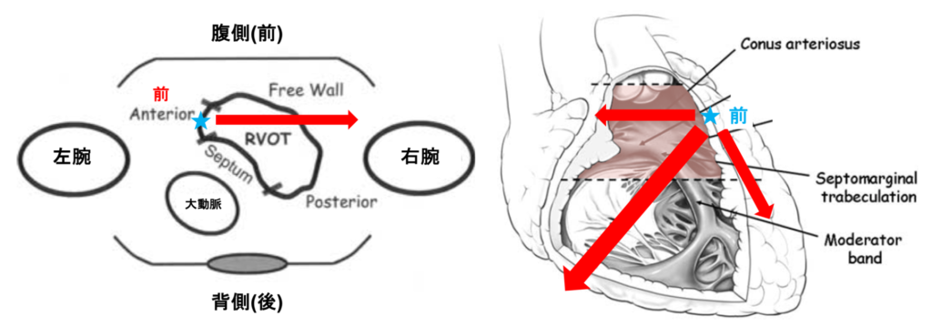
前壁からの興奮はaVL誘導と反対方向(Ⅲ誘導)に向かうため、aVL誘導でQ波(陰性成分)が大きくなります。
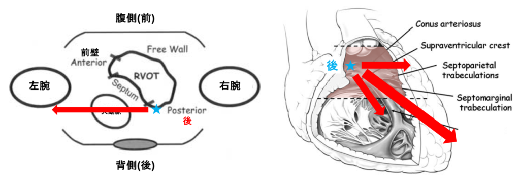
後壁からの興奮はaVR誘導と反対方向(Ⅱ誘導)に向かうため、aVR誘導でQ波(陰性成分)が大きくなります。
右室流出路自由壁
・中隔起源に比べ幅の広くて低いR波
・Ⅰ誘導で陽性成分が大きい※中隔側起源と対照的
・胸部誘導の移行帯はV3~V4以降(中隔側起源よりも時計回転方向になることが多い)
・aVL誘導とaVR誘導のQ波はaVR誘導のほうが深い※中隔側起源と対照的.
下壁誘導
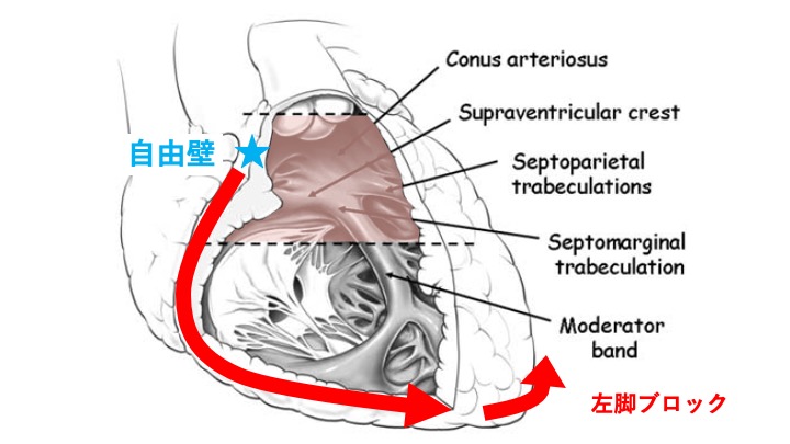
自由壁起源では右室の興奮が左室の興奮に先行します(phased excitation)4)。そのため、両心室の興奮のずれにより下壁誘導、I誘導に RR’パターン(R-R’間隔≧ 20ms)を認めます(R:右室の興奮、R’:左室の興奮)。また、右室と左室がバラバラに興奮することで起電力が分散されて下壁誘導のR波の波高は中隔起源に比べて小さくなります。
Ⅰ誘導
参考文献
1)Idiopathic left ventricular tachycardia. Catheter Ablation of Cardiac Arrhythmia- Basic Concepts and Clinical Applications-, ed by Wilber D, Packer DL, Stevenson WG, Blackwell Publishing, Oxford, p298~313
2)12誘導心電図波形を用いた特発性心室不整脈起源の診断. 心電図 2010; 30: 453-65.
3)循環器ジャーナル.2017 Vol. 65 no. 3
4)Prevalence and electrocardiographic characteristics of idiopathic ventricular arrhythmia originating in the free wall of the right ventricular outflow tract. Circ J 68 : 909-914,2004
深掘りしたい方へ
そのPVCはどこから-12誘導心電図からのアプローチ P10-21
起源推定に一点集中した名著。これ以外にお勧めする本がありません。もしあれば教えてください。書評に関しては下のリンクからお読みください。